Cart is empty
Home/Our Publications/Educational Articles/Cosmetic Tattooing & MRI’s - Diametric Particle Agitation Hypothesis (DPA)
Cosmetic Tattooing & MRI’s - Diametric Particle Agitation Hypothesis (DPA)
31/10/2010
by Andrea Darby - Master Medical Tattooist & Industry Educator
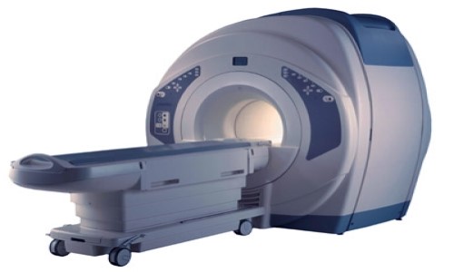
A Cosmetic Tattooist may encounter questions from clients about the possible effects on their tattoos when undergoing Magnetic Resonance Imaging tests (MRI), due to various media reports and internet publications that have referred to adverse effects.
This article will attempt to shed some light on the topic and presents the
various hypothesis's for the reports within the medical literature.
▼ Continue Reading ▼
|
Background MRI’s may be referred to variously as Magnetic Resonance Imaging (MRI), Nuclear Magnetic Resonance Imaging (NMRI), or Magnetic Resonance Tomography (MRT). An MRI is a medical imaging test designed to provide a computerised image of internal body structures which can also be printed onto a paper report or onto radiographer’s photographic film (an x-ray type image). Unlike an x-ray test the MRI machine does not use an ionizing radiation source to create the image instead the MRI uses extremely powerful rotating magnetic fields. By creating the rotating magnetic fields the scanner is able to compile cross sectional images of body structures which may provide an alternative diagnostic approach to other tests such as x-rays and ultrasounds. MRI of the Brain
You are most likely aware that compounds containing iron are strongly attracted to a magnetic field and that they can easily become magnetised, however most substances will produce a magnetic field in the presence of a strong magnetic source. This is what provides the MRI the ability to create an image of the body by scanning and recording the variations in the magnetic field when applied to the body structures.
Ferromagnetic / ferrimagnetic – These are the materials that you would probably recognise as being affected by a magnet they usually contain ferrous compounds (iron/iron oxides etc), the effect of a magnetic field can be physically felt and the material is capable of retaining magnetization.
Compounds within cosmetic tattoo pigments and body art inks are solutions that frequently contain iron oxides and a variety of other non ferrous metal oxides such as titanium dioxide as well as glycerine, water, and sometimes carbon and other compounds, in addition cosmetic tattoo pigments and body art inks have been found to contain nano particles both in the bottle and after implantation into the skin1. When first tattooed into the skin some of the water within the pigment would tend to be removed and pigment molecules become fixed into the upper level of the dermis by specialised cells called macrophages and fibroblasts. Over a period of 4-6 weeks the compounds within the tattoo pigment would tend to concentrate slightly as fluid is removed into the lymphatic system and the pigment would most likely remain in thick paste like consistency surrounded by the bodies fixating immune cells. Due to the particle agglomeration and the robustness of the colourants they prove difficult for macrophages to break down ultimately a compromise is reached and a fibrin rich encapsulation/matrix surrounds the pigment resulting in long term pigment fixation within the skin. In spite of the 'fixation' compromise by macrophages they continue with attempts to break down of tattoo pigments by producing hydrogen peroxide. When the tattoo pigment compounds are subjected to strong magnetic field, such as is applied by a MRI, theoretically there may be a range of magnetic properties within the tattoo pigments Ferromagnetic, Paramagnetic and Diamagnetic. The question of course is if the affect of the magnetic field on the pigment is likely to be any different to normal body tissue and if the affects are likely to be adverse effects.
The above researcher’s surveyed 1032 patients who had undergone MRI of those 135 had some type of permanent cosmetics prior to the MRI and only two of those (1.5%) experienced slight sensations of tingling and or burning, and in both cases the symptoms were only transient and it did not prevent the MRI from being performed.
William A. Wagle and Martin Smith reported in;
This of course suggests that interactions between MRI and tattoos do not necessarily just relate to pigments that contain iron oxides that are ferro/ferrimagnetic compounds resulting in an attraction to the strong magnetic fields, because in the above instance the tattoo pigments were shown to be nonferrous, however it is also possible that the certificate of analysis might not have been accurate. It is known that heavy metals such as lead and mercury are diamagnetic and lead can actually be made to levitate in the presence of a very strong magnetic field due to the strong repelling effect that can occur with some diamagnetic materials. An obvious question and concern in the case reported above is why compounds containing arsenic, lead, barium and mercury were in a tattoo pigment in the first place and this highlights the importance of sourcing pigments from reputable manufacturers who manufacture using safer ingredients and whom disclose their full ingredient lists. Digressing briefly; we note with some vague interest that the popular television program myth busters evaluated the unreferenced claim of exploding tattoos during MRI and their anecdotal testing results failed to produce any such effects. During our own empirical experience we have not had a client, nor been informed of about any cosmetic tattooist who has had a client, who has complained of adverse effects from undergoing an MRI that was attributed to their cosmetic tattoos. Within a presentation by Kasper K. Alsing at the European Congress on Tattoo and Pigment Research the author discussed the possibility that transient burning sensations during MRI scans in a small number of cases may be coincidental, research conducted at the The Tattoo Clinic, Bispebjerg Hospital Copenghagen Denmark found there was a minute but statistical significant increase in temperature in iron oxide pigments after subjecting them to MRI scan but not a sufficient increase to cause thermal skin burn, iron oxide pigments that were tested all contained metals with mixed magnetic properties. We note that the tattoo pigment was tested in vitro (not in vivo) which may affect the results but nonetheless the research was significant and the conclusion reached was
"It is a myth that magnetic resonance imaging causes thermal burn as noted in the medical literature" (sic in patients with cosmetic tattoos)2. Possible Cause of Burning Sensations (in isolated cases) During MRI Hypothesis: Diametric Particle Agitation (MRI-DPA)
and or Accelerated Fenton's Reaction ►
Reactive Oxygen Species (ROS). For example if various compounds within the tattoo pigment are ferro/ferrimagnetic and or paramagnetic (attracted by the magnetic field) and other compounds are diamagnetic (repelled by the magnetic field) then theoretically this may result in diametric agitation of the compounds within the skin and the production of heat resulting in cellular tissue damage either due to particle friction/rotation when under the influence of the strong magnetic fields of the MRI. Nano particles of tattoo pigment within the skin may be clustered within agglomerates (smaller particle chemically bound into larger particles) and the nano particles
within the agglomerates may also have differing magnetic properties. Hydrogen peroxide produced by macrophages could also create an accelerated Fenton's type reaction with iron oxide nano particles in the presence of a rotating magnetic field as the iron oxide nanoparticles absorb the energy from the magnetic field and convert it into heat via Brownian relaxation (physical rotation of the particles) and Neel relaxation (rotation of the magnetic moment)3
, the generation of heat in this way using magnetic fluid thermal therapy for
the treatment of cancer has been studied quite extensively4,5. Also Wydra et al. observed enhanced degradation of
methylene blue by free radicals generated by iron oxide nanoparticles heated in
an alternating magnetic field in the presence of hydrogen peroxide. In short there is abundant information within the medical and scientific literature to support
MRI Diametric Particle Agitation Hypothesis Pigments containing high concentrations or large particles of iron oxide compounds may potentially also cause issues due to simple ferro/ferrimagnetic attraction or RF-induced electrical currents, there has been some suggestion that older tattoo pigments were more susceptible due to higher concentrations of iron oxide compounds and therefore older tattoos may be more susceptible to adverse effects, however we have been unable to find any verifiable evidence for this claim. and or 2) Minor cellular tissue damage due to diametric/rotation forces forcing apart tattoo pigment agglomerates and or damaging cellular structures or the fixating fibrin encapsulation/matrix. and or 3) Accelerated Fenton's type reactions between iron oxide nano particles and hydrogen peroxide produced by macrophages in the presence of a rotating magnetic field causing the creation of reactive oxygen species (ROS). OR 3) RF-induced electrical currents resulting in tissue heating. OR 4) Other unidentified or unrelated causes (i.e. unrelated coincidence). The cellular tissue damage could be likened to the effects of slight frostbite (congelation) with the less severe cases of tissue damage resulting in transient reddening of the skin and the more severe cases resembling a burn perhaps with minor extravasation (oozing) of intracellular fluid contents caused by the creation of ROS. In the (hypothetical) illustration in Figure 3. below the particles within the pigment compound with differing magnetic properties, as depicted in black and grey in the illustration, are forced into diametrically opposite directions under the influence of the MRI magnetic field resulting
release of nano particles from their agglomerates, particle spin, surface
heating of the particle and possibly a Redox reaction or minor rupture of the fibrin encapsulation/matrix that was previously surrounding and fixating the pigment within the skin.
... are better models for describing the cause of; skin tingling/heating/burning/cellular rupture as opposed to simple ferromagnetic attraction. In contrast Creixell et al. and Polo-Corrales et al. demonstrated that surface heating of a nano particle can be induced without affecting the temperature of a surrounding solution, with as much as a 10 degree Celsius differential. 6,7 In a very small number of cases stronger reactions have occurred that resulted in injury to the patient with cosmetic tattoos, it is possible that traces of heavy metal compounds within the pigments may have played a part in those stronger reactions, possibly caused by DPA ► Redox or other unknown causes. At this stage the cause of tingling/burning sensations and skin effects in these small number of cases within the medical literature seems inconclusive and may require further investigation by the scientific experts to provide a definitive answer on this issue.
We would recommend that cosmetic tattooist always ensure that they source their pigments from reputable manufacturers who produce high quality pigments and provide detailed information about ingredient lists. Pigments containing compounds of arsenic, lead, barium, mercury and the like should not be used for cosmetic tattooing not only because of the potential MRI risks but also because of the potential general health effects that may result from tattooing those materials into the skin.
References
Copyright © 2010 CTshop.com.au & the article author All Rights Reserved. No copying, transmission or reproduction of site content is permitted without our prior written consent.
Printing Restriction: This article is print disabled, please read our Intellectual Property & Copyright Policies if you would like to request a copy or permission to use the article content for any purpose. |
Main Menu
- Eyeliner Tattooing vs Dry Eye
- MicroBlading - First Things First
- Cosmetic Tattoo Training Standards
- Carcinomas in Tattoos a Statistical Anomaly
- Lash or Brow Growth Enhancing Serums & Tattooing
- What Influences the Colour of a Cosmetic Tattoo?
- Hygiene Protocols Update : Surface Cleaning Wipes
- Preventing & Managing Disputes
- Warm vs Cool Colours
- Age of The Alpha Metrosexual
- Who Will Buy a Poorly Iced Cake?
- Australia now has a Board Certified MicroPigmentation Instructor
- Robot Tattooists?
- Postcards From Birmingham
- The SCAPP Scale - Personalising the Micropigmentation Service
- How to Choose Your PMU Artist
- Scalp MicroPigmentation - More Than Just Ugly Scars?
- Permanent Eyeliner - Avoiding Complications
- Personal Protective Equipment - Are You Covered?
- 3D Nipple Tattooing a New Service?
- Why Do Cosmetic Tattoos Change Colour? - (Part 1)
- Why Do Cosmetic Tattoos Change Colour? - (Part 2)
- Smart Tattoos Are They The Future?
- Presentation: Adding Cosmetic Tattoo to Your Salon
- Cell Phone Vibrating Tattoos
- UK Survey - One Third Regret Their Body Art Tattoo
- Collaborating & Consulting with Dr. Linda Dixon
- Stem Cell Research - Inside the Lab
- When Marketing Via News Media Goes Wrong
- Client Pre-Treatment Screening Questionnaire
- Permanent Makeup Google Search Trends
- Potential Causes of Nosocomial Type Infections in the Salon-Clinic Setting
- Topical Anaesthetics & Cosmetic Procedures
- Introduction to the Fundamentals of Colour Perception
- Clients With Unexplained Loss of Outer Eyebrow Hair
- Hyperpigmentary Skin Conditions & Cosmetic Tattooing
- Cosmetic Tattooing & MRI’s - Diametric Particle Agitation Hypothesis (DPA)
Site News Selection
Educational Article Selection
Regulatory Article Selection
Client Case Studies Selection
Science Library Selection
Complete regrowth of hair following scalp tattooing in a patient with alopecia universalis
31/01/2023
Atypical Intraepidermal Melanocytic Proliferation Masked by a Tattoo: Implications for Tattoo Artist
20/09/2018
Chemical conjunctivitis and diffuse lamellar keratitis after removal of eyelash extensions
26/08/2018
Scarless Breast Reconstruction: Indications and Techniques for Optimizing Aesthetic Outcomes
07/04/2018
High speed ink aggregates are ejected from tattoos during Q‐switched Nd:YAG laser treatments
28/03/2018
Unveiling skin macrophage dynamics explains both tattoo persistence and strenuous removal
08/03/2018
Granulomatous Tattoo reaction with Associated Uveitis successfully treated with methotrexate
08/02/2018
Identification of organic pigments in tattoo inks & permanent make-up using laser mass spectrometry
07/02/2018
Microbiological survey of commercial tattoo and permanent makeup inks available in the United States
03/02/2018


